Dear Nurses

Saturday, March 29, 2008

CRANIAL NERVES IN REVIEW


Updated 5/24
Dear nurses,

Stroke Series Assessment is no longer available. 

The largest of the cranial nerves, is the Trigeminal.
                 This nerve contains three branches:
                Opthalmic ( corneal blink reflex). 
                           Maxillary - which supplies the upper jaw
                   Mandibular - controls the lower jaw.
Learn more :Cranial nerves


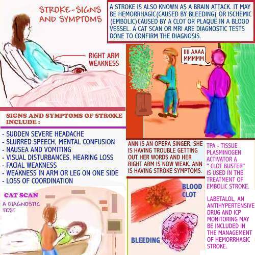
In the image above, Ann is an opera singer.
While on stage, she suddenly starts having
trouble getting her words out. Her right arm
also feels weak. These symptoms are classic
of stroke symptoms.

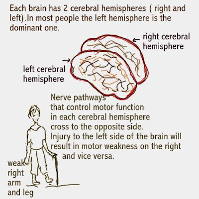
Severe headache, mental confusion, visual
disturbances and right or left sided weakness
ma occur.


      Learn more: The care plan
For more helpful information, please click on the links below:

CEREBRAL FUNCTION IN REVIEW


The human brain is made of 2 cerebral hemispheres,
right and left. The corpus callosum forms a bridge that
joins them together. Each hemisphere has four lobes
( frontal, parietal, temporal and occipital).

Millions of nerve cells called neurons originate in the
brain and transmit information all over the body. The
nerve pathways in the brain cross over. This results in
the left brain controlling the functions of the right and
vice versa.


Learn more about : Subarachnoid Hemorrhage


Learn more about : Aneurysms

Friday, March 21, 2008

HAPPY EASTER TO ALL NURSES


HAVE A GOOD EASTER WEEKEND! Enjoy learning more about the Clinical Setting.

FLORENCE NIGHTINGALE REMEMBERED


Hi guys,


Do you ever stop to remember how the Nursing profession got going? Well, let us not forget to give the credit to Florence Nightingale.Florence Nightingale. The one who started with simple things such as love, caring and compassion.
Today, we have come along way from the simplicity that started this noble profession, but those
words can still be valuable to us all. So, let us try to remember as we celebrate this Easter , to try to love and care for our patients, family and coworkers.
I trully appreciate all that I have learnt over the years, from bad situations to good ones in the Clinical Setting. These have all come together to make a contribution, that I can share with you.

Saturday, March 15, 2008

THE CLINICAL SETTING STEP BY STEP

Updated 12/24

Dear nurses,
The Clinical Setting Step by Step is no longer available.
New information is provided below.

Regardless of where a nurse works, doing good communication
skills, assessment tools and sound clinical judgment are all part
of a days work. Learn more: Here

ANEURYSMS IN REVIEW


Updated 2/25
An aneurysm is a bulge or balloon in a blood vessel.
The majority of aneurysms occur in the abdominal 
area.They may also occur along the blood vessels
that divide, after leaving the abdominal area.
An example of this phenomenon is the iliac artery.

PARALYTIC ILEUS IN REVIEW


Updated 6/23
Paralytic ileus may occur in the postoperative period.
Simply, click on the link:
and enjoy watching the video:

LIVER FAILURE IN REVIEW


Scenario: In the image above, Mr.O is in liver failure.
Past history includes 20 years of alcohol ingestion. His
liver is enlarged and his abdomen is grossly distended.

He is also lethargic and has pedal edema.
Learn more by clicking on the link:
Liver and renal failure

Learn more about Liver and kidney failure

Saturday, March 08, 2008

ADDRESSING ASYSTOLE AND OTHER LETHAL HEART RHYTHMS

Updated 7/25


The image above shows a Code blue in progress. This is 
happening in the emergency Room. Always follow your
Institution's policies and procedures for Code Blue. 

What is the role of the nurse who is assigned to the Code Blue?

The nurse has an important role in effective communication.
There are many responsibilities.
- staying calm in a chaotic situation
- giving clear instructions to coworkers
- ensuring that MD orders are conveyed correctly
- proper documentation of vital signs and all treatments /
  interventions that take place ( defibrillation for example)
  - the nurse becomes the " binding force.


Addressing lethal EKG rhythms

There are some EKG rhythms that are considered to be lethal.
Instant intervention is necessary for good outcomes. Here are
the most lethal rhythms:

Asystole - sometimes referred to as " flat-line. The electrical
heart's conduction system is not functioning. There are no
ventricular contractions. The patient is pulseless and
unresponsive. immediate action has to be taken. 

Ventricular Tachycardia - There may or may not be a pulse
present. The ventricles are beating rapidly and there is no atrial
activity. Blood flow to the vital organs is compromised.

Ventricular Fibrillation -  There is a chaotic rhythm and no
atrial activity. The ventricles are quivering , so there is no 
cardiac output. There is no pulse and the patient may become
unresponsive. Immediate action to defibrillate is usually done.

***American Heart Association has protocols in place for each 
EKG rhythm.

The postoperative patient is at risk for hypothermia.
Fluid loss and exposure in the surgical suite, may predispose
 to hypothermia. A lethal cardiac arrhythmia may result.

 Hypothermia increases the body's demand for oxygen
 and if left untreated can lead to a myocardial infarction
 (heart attack). Trauma resulting in excessive blood loss, 
 as well as alcohol ingestion, may also lead to hypothermia.

ADDRESSING PEA (Pulseless Electrical Activity)

Updated 7/25


 PEA is a cardiac arrhythmia that requires immediate treatment.
 ACLS guidelines are followed to treat this condition. Typically,
 patients have electrical conduction from the heart, but the clinical
 picture, is a patient who does not respond and needs CPR.

Causes of PEA include:
- Trauma, hypothermia
- Hypvolemia ( A common cause)
- Severe dehydration
- Electrolyte imbalances
- Pulmonary embolism
- Drug overdose
- Heart attack , cardiac tamponade and more

Treatment is directed at correcting the cause of the problem.
Drugs such as Epinephrine and Atropine are included in the 
treatment. ACLS protocols are followed.


Learn more about: Cardiopulmonary Arrest

CORRECT ASSESSMENT OF CHEST PAIN IN THE CLINICAL SETTING

The consequences of ignoring chest pain in the clinical setting may be fatal. Chest pain should never be ignored. Below, the result of the above nurse's decision is addressed.
For more information on assessing chest pain visit:
http://www.dearnurses.com/chest_pain_series_-_master_your_clinical
and
http://www.dearnurses.net/the_clinical_setting_step_by_step

WHAT IS WRONG WITH THIS EKG-VENTRICULAR FIBRILLATION

Ventricular Fibrillation is a lethal rhythm and requires immediate and skillful treatment. The ventricles of the heart "quiver" and there are no heart contractions. It is commonly seen in cardiac arrest.The patient above, was successfully converted to Sinus Rhythm with PVCs.

You may also enjoy watching the Clinical Videos called SESSIONS, in particular,
watch Sessions 16 through 18.

WHAT IS WRONG WITH EKG-TREATMENT OF VENTRICULAR FIBRILLATION

Updated 1/25
Dear nurses,
Dearnurses.com is no longer available.

Scenario:   
Ventricular Fibrillation is a lethal rhythm.
Its hallmark is no pulse, no heart contractions
and a chaotic EKG rhythm demonstrated above.
For more updated information, please click on the link:
Chest pain and the EKG

Causes and Treatment of Ventricular Fibrillation
are outlined above.


CORRECT CHEST PAIN ASSESSMENT


Chest pain ( Sessions 12) should never be ignored, no matter how simple. Assessment, documentation and intervention if necessary, should be done. These simple steps above
may be life-saving.
Enjoy reading Chest Pain Series for more information on this topic and
Myocardial Infarction(Chapter 1).

Saturday, March 01, 2008

HAVE A GOOD WEEK AND ENJOY LEARNING

Updated 7/25

Dear nurses,

There is no need to feel like you are drowning.
Below are new topics and links for learning.


Here are some diagnostic tests that are done frequently.
Follow the links below:
1. Chest X ray
2. CAT scan
3. EKG
4. MRI scan
5. Ultrasound
6. Echocardiogram

Learn more about : IV site care and assessment

   Learn more : The Nursing Process

WHAT IS WRONG WITH THIS EKG SERIES - TORSADES DE POINTES


Hi guys,


Hope you had a good week. I have tried to simplify TORSADES DE POINTES , which is a form of Ventricular Tachycardia. It is commonly found in patients who are chronic alcoholics and who are poorly nourished. Lack of Magnesium ( read also Preeclampsia, chapter 3)is usually the cause in chronic alcoholism.
Treatment is directed at correcting the cause.
For more information on Torsades de Pointes visit:
for Torsades go to page 33

You may also enjoy watching the Clinical Videos called SESSIONS, in particular
watch Sessions 16 through 18.

RECOGNIZING TORSADES DE POINTES

Dear nurses, 
See below for updated information.


Torsades de Pointes is a form of Ventricular Tachycardia. It is rare and
requires IMMEDIATE TREATMENT!
Causes include hypomagnesium, antiarrhythmic drugs that may prolong the Q-T interval and
myocardial infarction. Quinidine may cause Torsades de Pointes.
Symptoms include - Dizziness, chest pain, shortness of breath, hypotension and palpitations.


Dear nurses,
For more updated information, please
learn more by clicking on the link:
Updated 5/23

WHAT IS A PVC?

Updated 2/25

The image below, demonstrates chest pain brought
 on by stress. Mrs. K has an overly demanding boss.
 Not only is she experiencing chest pain related to stress,
 but her heart is having PVC's ( premature ventricular 
contractions), as reflected in the EKG.


A PVC is a premature ventricular contraction.
Typically, the ventricle contracts prematurely 
and the QRS is wider than normal. 

Heart disease may or may not be present. Anxiety/stress
are known causes. Other causes include:- 
Myocardial infarctionAmphetamines, Cocaine,
Caffeine and over-exhaustion. Treatment is
directed at the cause.Learn more:Chest pain and the EKG